Lymph node
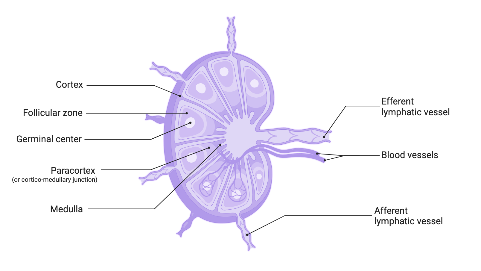
Lymph nodes are very small structures in their physiological state, 1 to 15 mm in diameter. The 500 to 1,000 lymph nodes in the body are linked together by lymphatic vessels to form lymph node chains which will drain into increasingly larger lymphatic networks, up to the thoracic duct where the lymph is reinjected into the systemic venous system. Lymph nodes are connected to both the blood system and the lymphatic network. Multiple lymphatic vessels enter the node while a single lymphatic efferent vessel exits, which tends to concentrate the lymphatic network as it is collected.
Structure
Lymph nodes are made up of three concentric zones, from the outside to the inside.
The cortex
It mainly contains B cells, macrophages, and ollicular dendritic cells. It is the place of B cell activation. It is organised into follicles:
- primary follicle, i.e. follicle without germinal centre
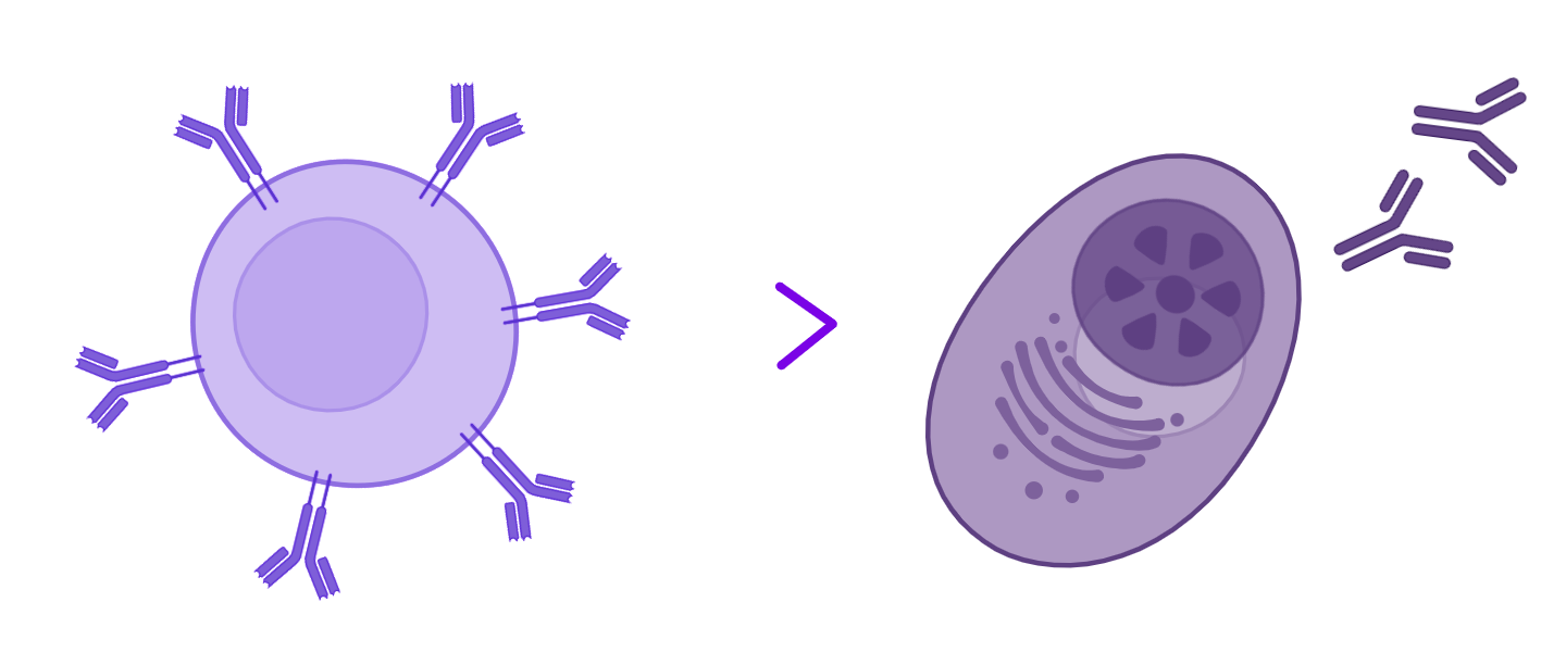
This is the site of the primary humoral immune response. It is within the primary follicles that short-lived plasma cells are generated, producing IgM with low affinity for the antigen. - secondary follicle, i.e. follicle with germinal centre
This is the site of the secondary humoral immune response. Within the germinal centre, B cells will undergo somatic hypermutation of their BCR, and possibly class switching (see B cell activation).
If this hypermutation increases the affinity for the antigen, then the B cells will differentiate into memory B cells or long-lived plasma cells producing IgG, IgA, or IgE. Newly formed plasma cells can then:
- either migrate towards the medulla and secrete immunoglobulins which then pass into the blood;
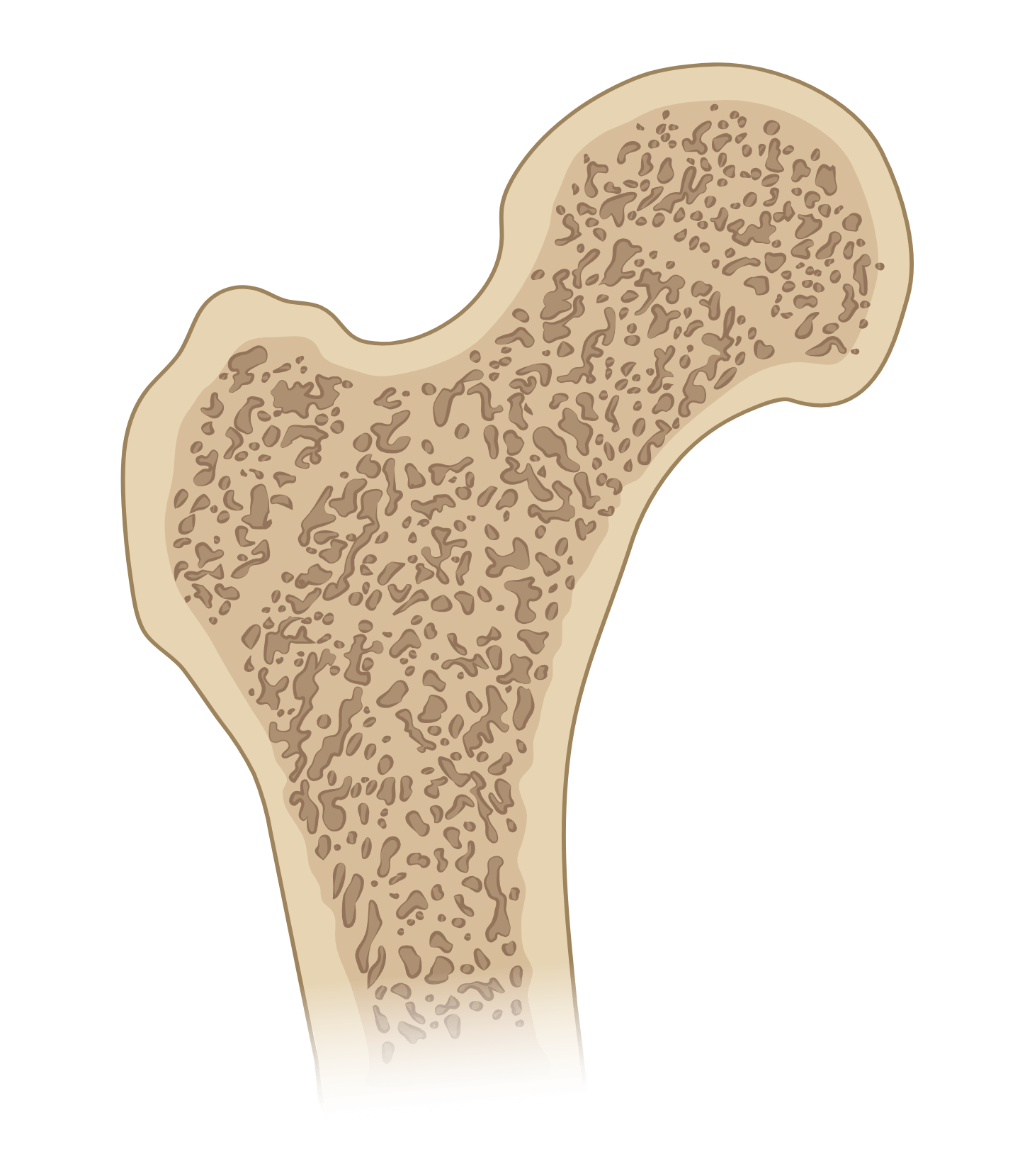
- or exit through the efferent vessels, join the blood circulation before relocating in the bone marrow, also to secrete immunoglobulins.
Paracortex (or cortico-medullary junction)
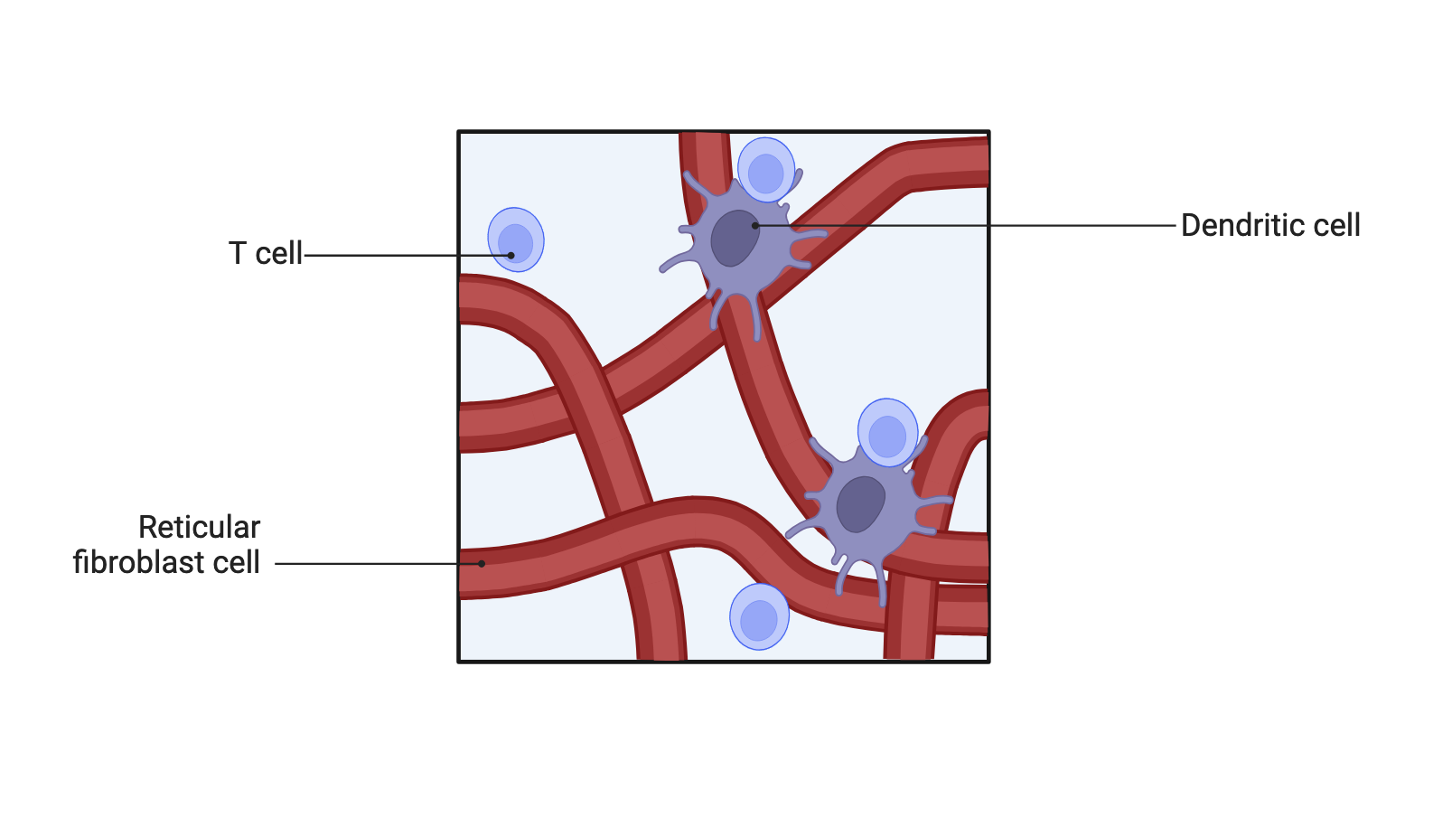
It mostly contains T cells and dendritic cells, which have migrated from peripheral tissues to the lymph node. This is the site of activation of naive T cells by dendritic cells.
Medulla
The medulla corresponds to the place of passage of lymphocytes which leave the lymph nodes via the efferent lymphatic vessels.
Main functions
Lymph node is organised to promote the encounter between antigens and naive lymphocytes, then the activation of lymphocytes if necessary.
Antigens arrive in the lymph node through the afferent lymphatic vessels:
- either processed by a professional antigen-presenting cell: they are then embedded in a Class I or Class II HLA molecule.
- or in particulate form: they can then be taken care of by professional antigen-presenting cells which are present in large quantities in the lymph node.
For their part, naive T and B cells arrive in the cortical zone of the lymph node via HEVs (High Endothelial Venules).
- T cells are directed to the paracortex to interact with dendritic cells, in order to find the [HLA-peptide] complex capable of activating them. T cells are guided in this pathway by the network formed by the cytoplasmic extensions of FRC (fibroblastic reticular cells), by the synthesis of adhesion molecules on their surface and by chemokine gradients.
- B cells remain at the cortex, they are directed by follicular dendritic cells, which play a major role in maintaining the structure of follicles and germinal centres, as well as in the presentation of antigens to B cells.
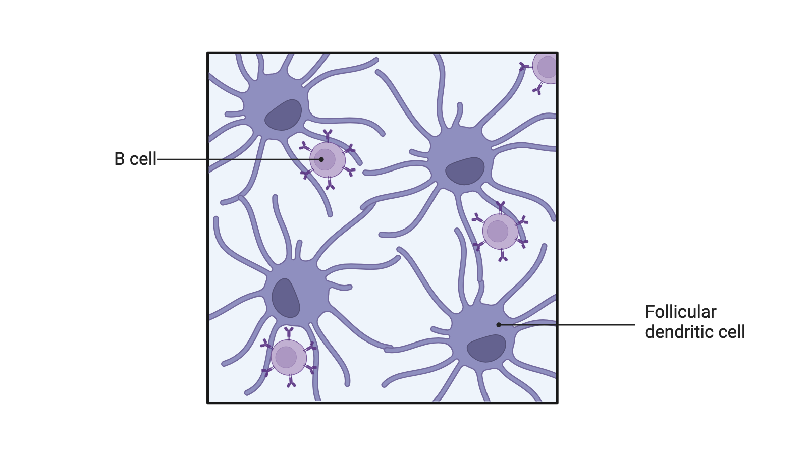
Attention to “false friends”: do not confuse dendritic cells with follicular dendritic cells
Follicular dendritic cells have this name because they are cells with large cytoplasmic extensions, located within lymphoid follicles.
Although they have the same name as dendritic cells, their role and characteristics are very different.
What needs to be remembered
Lymph nodes are organised to potentiate their two main functions:
- maximise the chances of encounter between the lymphocytes (T or B cells) and the antigen which can be recognized by these lymphocytes.
- if this encounter takes place, allow the activation of lymphocytes (T or B cells), so that they can exercise their functions, cytotoxic or auxiliary (for CD8+ and CD4+ T cells, respectively) or secretion of antibodies (B cells differentiated into plasma cells).
